

Congenital cardiac surgery · Cardiovascular tissue engineering
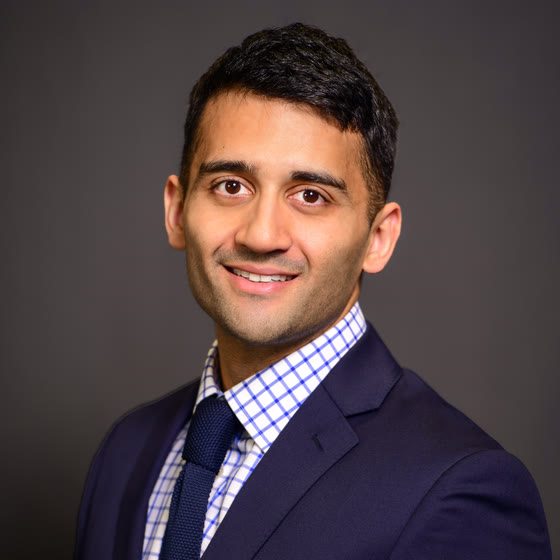
Syed Faaz Ashraf
Clinical PGY-6 integrated cardiothoracic surgery resident at the University of Pittsburgh. Three years of NIH-funded research in cardiovascular tissue engineering at Carnegie Mellon, building conduits, instruments and software for congenital heart disease.
Conduits and tissue
4 projectsA Fontan conduit that pumps
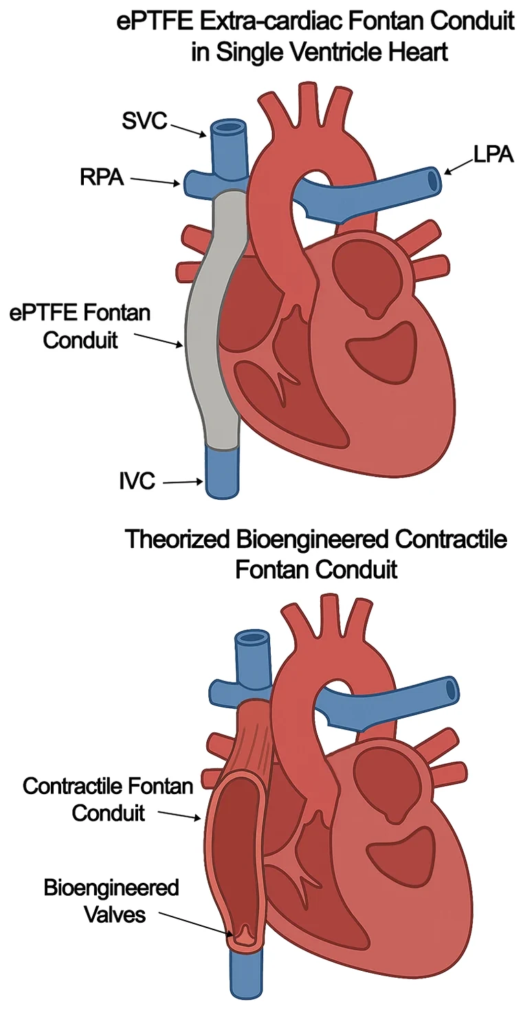
Patients with single-ventricle disease carry the long-term morbidity of a Fontan circulation because there is no sub-pulmonary ventricle. The aim is a pulsatile conduit of engineered heart tissue, acting as a neo-ventricle and generating 5–10 mmHg of subpulmonic pressure. To our knowledge it is the first bioengineered ‘organ’ to generate pulsatile pressure in vivo.
- Problem
- A Fontan circulation leaves no sub-pulmonary ventricle. Pulmonary flow is passive, driven by systemic venous pressure, and the ePTFE conduit that carries it neither contracts nor grows with the child.
- What I built
- Mouse-scale collagen conduits, FRESH 3D-bioprinted at three stiffnesses and cellularised with human stem-cell-derived cardiomyocytes and cardiac fibroblasts. They were matured in a custom bioreactor, then anastomosed end-to-end onto the infra-renal IVC of SCID/beige mice.
- Evidence
- 32 conduits anastomosed. They stayed patent and contractile up to a year in vivo, generating 8–10 mmHg of pulsatile pressure at six months. The collagen remodelled into vascularised neo-tissue, and the conduits captured on field stimulation at 1 and 2 Hz.
- Status
- Manuscript in writing, for submission to Science Translational Medicine. President's Award Paper, Congenital Heart Surgeons' Society 2025. Lillehei Award finalist, AATS 2025. AHA Scientific Sessions 2024 (Circulation. 2024;150(Suppl 1):A4144448).
- With
- Feinberg Lab, Carnegie Mellon University. Ashraf SF, Bliley J, Yi T, Feinberg AW.
- Funding
- NIH/NHLBI F32HL165847 — Principal Investigator, 2022–2024, $145,616. Additional Ventures Cures Collaborative.
CardioConduitNet
A deep-learning pipeline I wrote and trained. It segments a contractile engineered conduit on ultrasound (lumen, conduit wall, aorta), separates respiratory from cardiac motion, and reports regional contractility. Clinical echo software can't read anatomy like this, and the tissue-engineered hearts that follow won't resemble a human heart either.

- Problem
- Clinical echo software isn't built to read a contractile engineered conduit, so lumen, tissue and motion have to be traced by hand. The engineered hearts and pumping ‘organs’ that follow won't look like a human heart at first either.
- What I built
- I built this end to end, from the training data through to validation. The model reads ultrasound of the conduit, outlines lumen, wall and aorta automatically, separates breathing from heartbeat, and reports contractility segment by segment using the AHA wall-motion model.
- Evidence
- Processes 5,000 ultrasound frames in about two minutes, work that takes hours by hand. Automated outlines match hand-drawn ones closely (median Dice 0.85 lumen, 0.78 tissue), across 162 recordings 0.5–5.5 months after implantation.
- Status
- Manuscript in writing, for submission to Science Translational Medicine.
- With
- Regenerative Biomaterials & Therapeutics Group, Carnegie Mellon University
- Funding
- Additional Ventures Cures Collaborative. NIH/NHLBI F32HL165847 — Principal Investigator, 2022–2024, $145,616.
Printing the fibre architecture of the ventricle
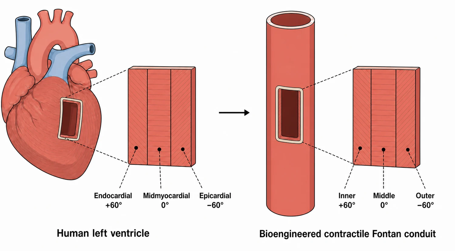
Heart muscle isn't laid down evenly. The fibre angle turns as you move through the wall, from about +60° on the inside to −60° on the outside, and that twist is what lets a ventricle wring rather than just squeeze. Tissue printed without it only shortens. If a conduit is going to pump, it needs the same architecture.
- Problem
- The conduits built so far contract, but their myocytes are laid down with no preferred orientation. In native myocardium the helix angle rotates from about +60° to −60° across the wall (Streeter, Circ Res 1969). Without that gradient a construct shortens but can't generate torsion.
- What I built
- A multi-layer print that lays each wall at a controlled helix angle: +60° inner, 0° mid-wall, −60° outer, mirroring the subendocardial, midmyocardial and subepicardial layers of the left ventricle. Fluorescent Carbopol makes every layer and its orientation directly visible.
- Evidence
- Current work in progress. Fibre-angle and contractility quantification is underway. The printing approach itself is demonstrated layer by layer in fluorescent Carbopol.
- Status
- Current work in progress.
- With
- Amanda Kang, PhD student at Carnegie Mellon, whom I mentor on this work; I designed and built the five-axis printer hardware it requires. Feinberg Lab, Carnegie Mellon University.
- Funding
- Additional Ventures Cures Collaborative.
A bioprinted valved conduit
Conduit choice for the right ventricle to pulmonary artery connection is poor, and valved conduits are worse. Nothing available grows with the child, so every patient comes back for reoperation. This is a bioprinted collagen conduit with a valve inside it, tested in a heterotopic heart transplant model in mice.
- Problem
- No RV-PA or valved conduit in use today grows with the child, so each one commits the patient to reoperation.
- What I built
- A FRESH-bioprinted collagen conduit with a valve printed inside it, anastomosed from the right ventricle to the left atrium in a mouse heterotopic heart transplant model, and perfused by the transplanted heart.
- Evidence
- Direct imaging of the valve opening and closing. Echocardiography shows the leaflet in diastole and systole, and colour Doppler confirms directional flow through the conduit.
- Status
- Ongoing work.
- With
- Jaci Bliley, Carnegie Mellon, and Christopher Breuer's group, Nationwide Children's Hospital.
- Funding
- Additional Ventures Cures Collaborative.
Instruments and software
3 projectsReplistruder 4.5 and the printer
Replistruder 4.5 and a custom bioprinter, built in-house as a successor to the Replistruder 4, the most widely adopted open-source syringe-pump extruder design in bioprinting.
- Problem
- FRESH bioprinting needs a syringe extruder precise enough for embedded printing, and the lab builds and adapts its own rather than buying them.
- What I built
- Replistruder 4.5, designed and built from the ground up as the successor to the Replistruder 4 (Tashman, Shiwarski, Feinberg; HardwareX, 2020), the most widely adopted open-source syringe extruder in bioprinting. Made in 3D-printer and CNC variants alongside a custom bioprinter on open-source Duet electronics, and released open source for any lab to build.
- Evidence
- $90 per extruder, 20% narrower than the Replistruder 4, with independent Z control on each so the Z offset between nozzles is eliminated. Prints collagen filaments consistently at 90 µm.
- Status
- Manuscript in writing, for submission to Scientific Reports (Nature Portfolio).
- With
- Feinberg Lab, Carnegie Mellon
- Funding
- NIH/NHLBI F32HL165847 — Principal Investigator, 2022–2024, $145,616.
ALINr
A low-cost, open-source camera system that finds each bioprinter needle by computer vision and calibrates the offsets between them, so multi-material prints land where they're meant to. I designed and built it, hardware and software, and released it open source.
- Problem
- Multi-nozzle bioprinting only works if the XYZ offsets between extruders are known. Touching each nozzle onto a marker on the bed risks contaminating the bioink and leaves ±100 µm of error; a laser micrometer costs upwards of $20,000.
- What I built
- ALINr, the Automated Lens-based Initial Needle Registration system. Cameras locate each extruder needle in X, Y and Z by detecting its outer diameter, and the printer calculates tool offsets from the first nozzle so every material prints in register. It exists in two forms: a camera carried on a collaborative robot arm, and a battery-powered Raspberry Pi unit that works inside a culture hood for sterile calibration.
- Evidence
- $280 in parts. Calibrating three extruders left mean errors of 52 ± 29 µm in X and 36 ± 26 µm in Y, a 3.5-fold reduction in X and 8-fold in Y and Z against point-based alignment, measured on three-nozzle collagen prints by optical coherence tomography.
- Status
- Manuscript in writing, for submission to Scientific Reports (Nature Portfolio). Poster, TERMIS World Congress, Seattle, June 2024. Published abstract: Tissue Eng Part A. 2024;30(15-16):S412.
- With
- Feinberg Lab, Carnegie Mellon University. Ashraf SF, Shiwarski DJ, PereiraTavares A, Tashman JW, Feinberg AW.
- Funding
- NIH/NHLBI F32HL165847 — Principal Investigator, 2022–2024, $145,616.
photoFRESH light-pipe
A fibre-optic light-pipe that delivers light into embedded 3D printing for layer-by-layer control of light-driven chemistry (photochemistry), developed as part of a team effort led by Dikyol C, O'Brien WB and Stang MA.
- Problem
- Embedded 3D printing lacked a way to apply localised light-driven chemistry (photochemistry) layer by layer without compromising print fidelity.
- What I built
- Multi-modal photoFRESH, a fibre-optic light-pipe that brings light-driven chemistry into embedded printing, giving layer-by-layer control of crosslinking, stiffness and attached biomolecules without disturbing living cells. I designed and built all of the hardware that makes it work.
- Evidence
- Demonstrated across the experiments reported in the preprint: photocuring inside the support bath, multi-material multi-wavelength printing, and biomolecule tethering with print fidelity preserved. Manuscript submitted to Science.
- Status
- Preprint: Dikyol C, O'Brien WB, Stang MA, Ashraf SF, et al. bioRxiv 2026.07.19.738036, doi:10.64898/2026.07.19.738036. Submitted to Science.
- With
- I designed and built all of the hardware and robotic gantry systems that make this technology work. Team led by Dikyol C, O'Brien WB and Stang MA.
- Funding
- Additional Ventures Cures Collaborative.
Teaching and service
2 projectsA calf thorax model for robotic IMA harvest
A calf thorax simulation model for robotic internal mammary artery harvest and endoscopic coronary artery bypass grafting on the da Vinci Xi. It was validated and published in Innovations, and it's now the default model in the curriculum for the STS robotic cardiac surgery workshops, held every other year nationally.
- Problem
- Robotic internal mammary artery harvest is the hardest part of endoscopic coronary bypass to learn, and there was no affordable, high-fidelity way to practise it. Commercial simulators are expensive and poor in tissue realism; the alternative is a live animal.
- What I built
- A calf thorax model for robotic internal mammary harvest and endoscopic coronary bypass on the da Vinci Xi, developed for the STS Workshop on Robotic Cardiac Surgery in Atlanta. I built the model and ran the one-day coronary session in 2022, then returned as faculty for the two-day session in 2024.
- Evidence
- Now the default model in the curriculum for the STS robotic cardiac surgery workshops, held every other year nationally: 50 models built per course, 80 cardiac surgeons trained each time.
- Status
- Published: Ashraf SF, Seese L, Hasan IS, et al. Innovations. 2024;19(6):633-639. PMID 39473044. First author. Podium presentation, STS Annual Meeting, San Antonio, January 2024.
- With
- Johannes Bonatti, UPMC, with Seese L, Hasan IS, Babu AN, Balkhy HH, Kiaii BB, Guy TS, Kaczorowski DJ.
- Funding
- NIH/NHLBI F32HL165847 — Principal Investigator, 2022–2024, $145,616.
TSRA
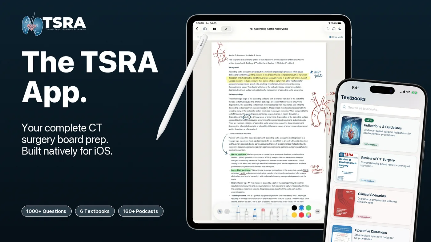
Founder and developer of the TSRA App, the official iOS study platform of the Thoracic Surgery Residents Association; Editor-in-Chief of TSRA Education Articles, Congenital Section Editor for the TSRA Indications & Guidelines volume, and author of three TSRA book chapters.
- Problem
- Trainees prepare for boards from six separate TSRA textbooks, a question bank and a podcast library, with no common home and no way to study offline between cases.
- What I built
- The official iOS study platform of the Thoracic Surgery Residents Association, which I built on my own and still maintain: six TSRA textbooks, 1,000+ board questions and 120+ podcasts, with flashcards, annotation and offline study. Published February 2026. I'm also webmaster of tsraweb.com.
- Evidence
- 1,500+ downloads and 600 paid subscribers at $60/year, about $36,000 a year in recurring revenue supporting TSRA education.
- Status
- Founder and Editor-in-Chief, TSRA Education Articles. Congenital Section Editor, TSRA Indications and Guidelines in Cardiothoracic Surgery (2026). Author of three TSRA book chapters. TSRA President-Elect, 2027–2028.
- With
- Thoracic Surgery Residents Association
- Funding
- Thoracic Surgery Directors Association; the app is otherwise self-funded through subscription sales.
- See it
- App Storetsraweb.com
01 FUNDING
- NIH/NHLBI F32HL165847 — 3D Bioprinted Collagen Vascular Conduits for Use in Patients with Congenital Heart Defects (Principal Investigator; $145,616)09/2022–08/2024
- Burroughs Wellcome Fund — Physician-Scientist Incubator Program Scholar, University of Pittsburgh2022–2024
- NIH/NHLBI T32HL160526-01 — Cardiothoracic Surgery Research Training Program
- Beckwith Frontline Innovation Grant — Modular Lung Transplant Surgical Anastomosis Simulator (Project Lead; $9,000)2022–2024
02 AWARDS
- Congenital Heart Surgeons' Society President's Award2025
- Congenital Heart Surgeons' Society, Best Resident Presentation Award2025
- AATS Lillehei Award, finalist2025
- American Heart Association, Genomic and Precision Medicine Travel Award2025
- TSDA Cardiothoracic In-Training Examination Prize, national top 10%2019–2020
03 SERVICE
- TSRA — President-Elect2027–2028
- TSRA — Secretary2026–2027
- TSRA — Editor-in-Chief, TSRA Education Articles2025–Present
- TSRA — Projects Chair2025–2026
- TSRA — Education Chair2024–2025
- TSRA — Executive Board Member2022–2024
- TSRA — Congenital Section Editor, TSRA Indications and Guidelines2026
04 SELECTED PUBLICATIONS
- Ashraf SF, et al. The cone repair allows right ventricle rehabilitation with excellent tricuspid valve function following the Starnes procedure. J Thorac Cardiovasc Surg. 2025;169(2):354-361.e3
- Ashraf SF, et al. Ebstein anomaly variant with anterior leaflet displacement. JTCVS Tech. 2024;28:120-123
- Ashraf SF, et al. Impact of the 2018 change in US allocation policy on adults with congenital heart disease. J Heart Lung Transplant. 2022;41(3):373-381
- Ashraf SF, et al. Development and validation of a low-cost, high-fidelity simulation model for robotic internal mammary artery harvest using the da Vinci Xi robot. Innovations. 2024;19(6):633-639
- Ashraf SF, et al. Predicting benign, preinvasive, and invasive lung nodules on computed tomography scans using machine learning. J Thorac Cardiovasc Surg. 2022;163(4):1496-1505.e10
- Bakirci E, Asghari Adib A, Ashraf SF, Feinberg AW. Advancing extrusion-based embedded 3D bioprinting. Biofabrication. 2025;17(2):023002
Clinical PGY-6 integrated cardiothoracic surgery resident at the University of Pittsburgh. Applying for fellowship positions in congenital cardiac surgery.
[email protected]